


In our group we recognize an urgent need to keep developing new tools, techniques and methods to study collagen, other proteins and composite materials that humans are made of at the tissue/cellular level without disrupting their fragile 3D architectures.
To that end, we custom- designed, built and maintain at UCR a cost-effective non-linear optical microscopy imaging and spectroscopy resource (Laboratory.html - Multiphoton Microscopy Imaging System). The existing commercial designs were adapted to obtain the optimal performance for the materials (including tissues) we worked with so far. This resource is unique to the Bourns College of Engineering (BCOE) and UCR. Our modular design is configurable to study different specimens under diverse settings and we use the light provided by a tunable laser device to instantaneously study structure and processes at very small scales (on the order of microns and smaller) within collagen hydrogel materials and other more complex biological systems and living organisms.
 |
 |
Polarization Second Harmonic Generation (SHG) and Two-photon Fluorescence (TPF) Imaging
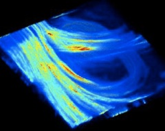
Polarization-based SHG and TPF microscopy can be used to follow development of the supramolecular structure of collagen within tissues and materials and possibly can separate normal collagen tissues from diseased.
An example of polarization-based SHG imaging separating collagen fibers as a function of the incident polarization. Similar polarization-dependent behaviour of SHG was previously described by other investigators on somewhat similar biological samples. |
(A) Horizontal polarization of laser excitation and horizontal orientation of analyzing polarizer (parallel to sample surface) selected fibers running parallel in x-y plane; (B) Circular polarization of laser excitation and horizontal orientation of analyzing polarizer (parallel to sample surface) with fibers visualized in (A) being subtracted to highlight fibers running in the plane other then x-y. (C) Merged image of (A) and (B). Proc. SPIE 9540-36 (2015) |
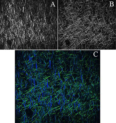
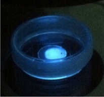
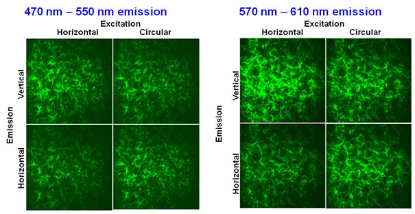
Polarization-based TPF imaging of genipin-modified collagen hydrogels shows that the intensity of fluorescence is at its minimum when excitation is horizontally polarized and orientation of analyzing polarizer is horizontal (parallel to sample surface). This observation is indicative of possible orientational differences in the conjugated moieties introduced as a result of genipin reacting with intermolecular and intramolecular collagen residues packed into fibril and fiber structures within hydrogels. |
Polarization selection of TPF of newly induced fluorescent collagen fibers. The levels of SHG signal are undetectably low and the SHG images therefore are not shown. Images are about 180 um x 180 um. Proc. SPIE 9540-36 (2015) |
Due to our unconventional approach to optically record collagen and cellular properties at the tissue level in a non-invasive manner, this research has a very high impact on the scientific community at large.
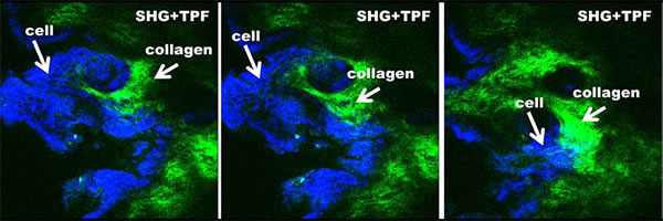
Multiphoton Microscopy (MPM) non-linear optical imaging of skin
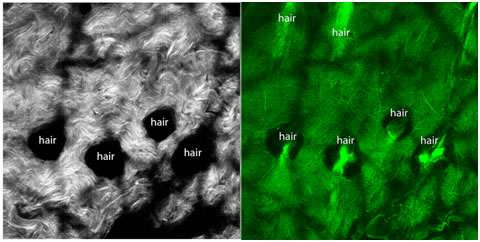
| Second harmonic generation (SHG) and two photon fluorescence (TPF) signals of MPM are used to visualize the structure of a completely intact mouse skin. In-situ images of the dermis of the skin show assembled collagen fibers that generate very strong second harmonic generation (SHG) signals (left). Two photon fluorescence (TPF) (right) aids in identifying hair follicle cells, elasitic and collagen fibers. No exogenous contrasts were used to obtain the images below. |
 |
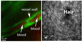
MPM has a direct relevance to studies of fundamental mechanisms underlying normal hair follicle physiology, development of its diseases, and in evaluating the efficacy of administered treatments. We can image deep into hair follicles in situ without mechanical sectioning of tissues or applications of exogenous dyes. |
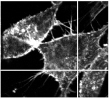
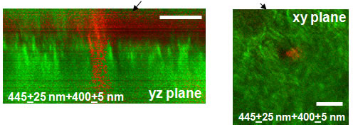
Left: Transverse optical section through a 3D MPM image ; Right: En face optical section at ~56 um through a 3-D stack in a 3D MPM image. Green: second harmonic signals (SHG) generated by extracellular matrix (collagen). Red: cellular fluorescence (TPF). Scale bar is 50 um. Journal of Biomedical Optics 12, 044003 (2007).
High resolution en face optical sections through a hair follicle:
Green: second harmonic signals (SHG) generated by extracellular matrix (collagen). Red: cellular fluorescence (TPF).
|

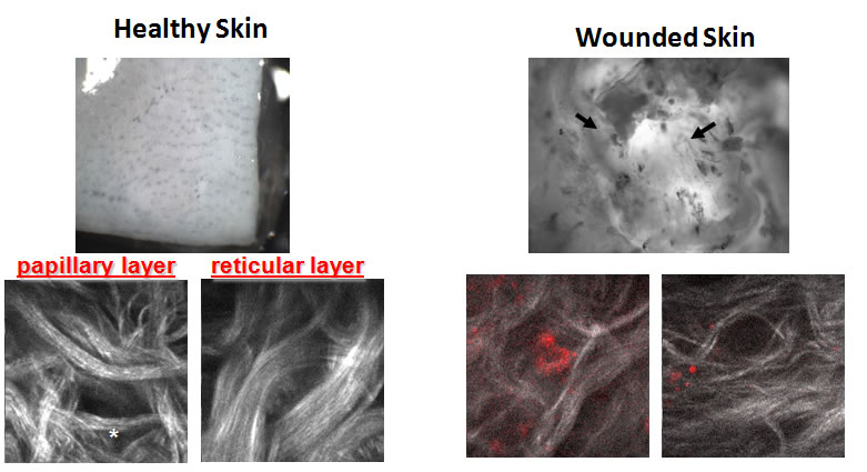
| MPM clearly distinguishes normal and pathological dermis therefore being relevant to skin research (wounds, ulcers, etc.) and skin-related product development. Proc. SPIE 6859, 1-9. doi: 10.1117/12.762702 (2008), Invited Paper |
Optical "section" of the human dermis (contrast: SHG from collagen)
|
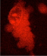
Modification of an epithelized tissue (skin, cornea,etc.) with cross-linking reagents for the purpose of strengthening of those tissues in diseases like for example keratoconus, highlights a critical need for the techniques such as MPM that can monitor the extent of tissue modification. For example, we find that the epidermis in skin and epithelium in cornea will protect collagen from cross-linking with glyceraldehyde, implying that strengthening will differ based on the cross-linker dose that is administered and exact delivery methods. Proc. SPIE 8587, 858725, doi:10.1117/12.2012305 (2013) |
Blue: TPF from cells cross-linked with glyceraldehyde; green: SHG of collagen that did not become cross-linked |
Our experitse in tissue imaging was very useful in identifying various features of wounds in collaboration with Prof. Martins-Green at UC Riverside: Wound Repair and Regeneration 20, 353-366 (2012) Journal of Diabetes Research vol. 2014, Article ID 562625 (2014) Clinical science DOI: 10.1042/CS20150393 (2015) and subsequent assessment of burns in live rats. |
Blood vessel (left) and neo-epithelium cellular structures (right) are developing within the regenerating skin of a live rat |
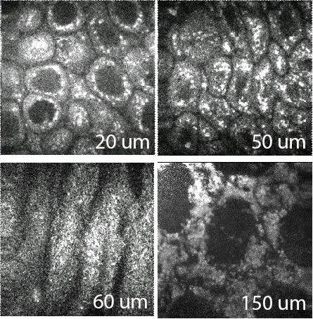
Multiphoton Microscopy (MPM) non-linear optical imaging of the eye
The high-resolution, epi-reflection MPM produces very selective signals from epithelial, stromal and endothelial layers of corneas. Second harmonic generation (SHG) and two photon fluorescence (TPF) signals of MPM of mouse cornea (x-y scans) show: a) the changing spatial organization of cells within the epithelial layer (first 50 um); b) collagen within stroma at 60 um;c) endothelial layer at 150 um. |
Journal of Biomedical Optics 11, 014013 (2006)
3D reconstruction of the mouse cornea from MPM images
|
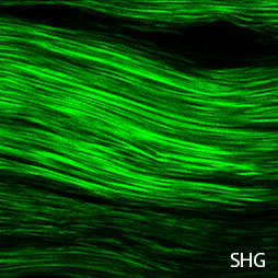
Multiphoton Microscopy (MPM) non-linear optical imaging of the tendons
Tendon is a fibrous connective tissue connecting muscles to bones and works together with bones to move them. Tendons can transmit tension due to a unique mostly parallel arrangement of collagen fibers within them. Tendon tears are thought to be due to weakening of collagen structures from overuse of tissues and excessive tissue strains.
Exogenous cross-linking of collagen is being looked into as a possibility to treat small lesions and tears in tendons to avoid acute injuries and invasive surgical repairs and to potentially benefit unmet clinical needs.
Second Harmonic Generation (SHG) signals from collagen fibres in rat tail tendons
|
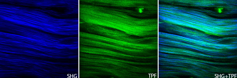
Second harmonic generation (SHG) and two photon fluorescence (TPF) signals of MPM in rat tail tendons strengthened with glyceraldehyde.
|
Copyright 2007 J.G.L. All rights reserved.


Extra Links: